Orthopaedics,
made clearer.
Anatomy. In a new dimension.
Interactive bone anatomy, orthopaedic planning and digital healthcare. Explore the simulators, discover clinical systems, and follow my academic work.
Explore the projects ↗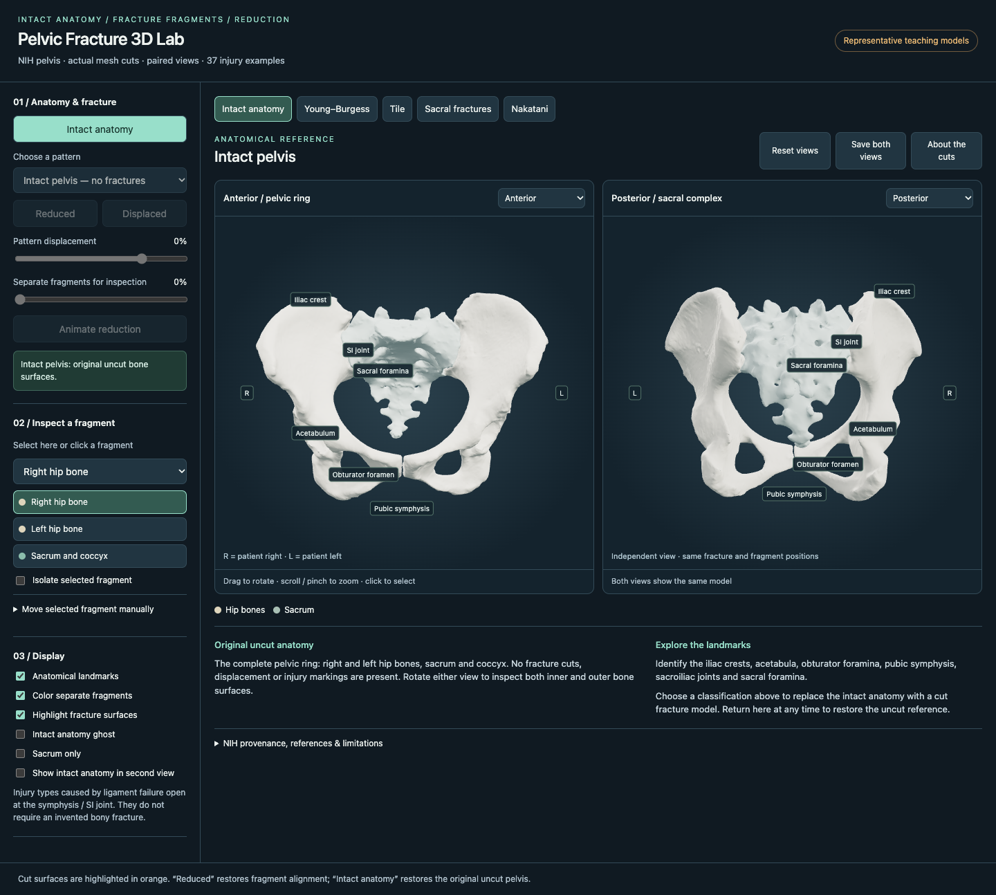
Structure. Stability. Movement.↗
Explore the whole picture.
Orthopaedic teaching, clinical workflows
and academic work, together.
3D Bone Lab ↗
Ten projects exploring fractures, positioning, imaging and reconstruction.
02 / PLANNING & MEASUREMENTPlanning Software ↗
OrthoPlan Studio and Bone Planning Studio. Launch the browser prototypes.
03 / DIGITAL HEALTH SYSTEMSClinical Systems ↗
Patient journeys, hospital architecture and AI-supported workflow projects.
04 / EVERYDAY WORKFLOWSClinical Digital Tools ↗
Explore the existing tools for doctors, nurses, research and documentation.
Find your way, one stage at a time ↗
Explore the IGIMS campus in 3D, select buildings and switch to the master-plan view. Indoor navigation awaits verified floor plans.
Open the hospital navigator ↗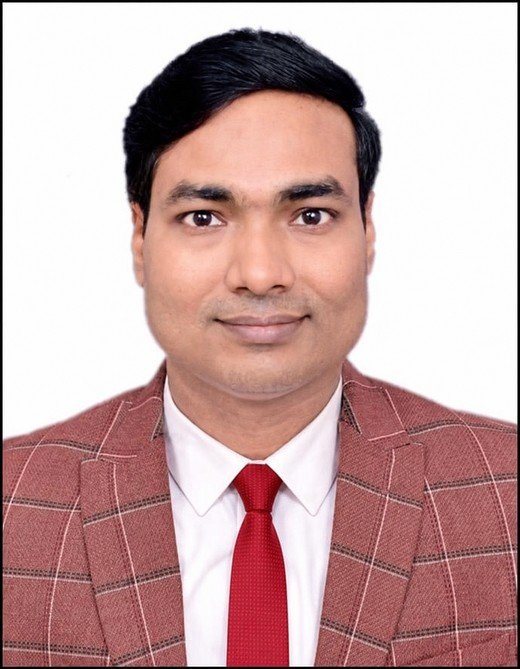
Dr. Rakesh Kumar
Associate Professor of Orthopaedics · IGIMS Patna
Academic profile, publications, research & courses ↗Your reports.
On the doctor’s screen.
A QR code connects the patient’s phone to the doctor’s browser. Share PDF reports and photos in a live session, with English and Hindi instructions.
Receive & save to computer ↗
Doctors: open this tool to start.
Patients: scan the doctor’s QR code.
Internet required on both devices.
Explore a different angle.
Ten projects. From understanding a fracture
to seeing beyond the image.
Built to explore.
Designed to understand.
Bone Atlas brings orthopaedic projects together in one place. Rotate anatomy, inspect fragments and explore how imaging connects to three-dimensional form.
Explore the simulators directly in your browser. Each project retains its own anatomy sources, credits and limitations.
Find your next perspective ↗Move beyond a static image.
Explore spatial relationships from your own viewpoint.
From image to anatomy.
Bring radiographic views and 3D models into the same collection.
Follow your curiosity.
Navigate by anatomical region or reconstruction workflow.